The Irish Wolfhound should be a strong, athletic dog with the personality of a sweet, easy-going couch potato. However, to maintain health and condition, he should not spend all his time lounging around the house. Daily walks, especially if you have a safe area for some off leash exploring are good for both body and soul.This allows the dog to exercise his muscles and his mind.To maintain good health, proper nutrition and proper exercise are key, but there are some medical issues that may arise in spite of your best efforts. I will discuss some of the more common ones here. For more information about IW health concerns I refer you to the national club
First, potential puppy problems:
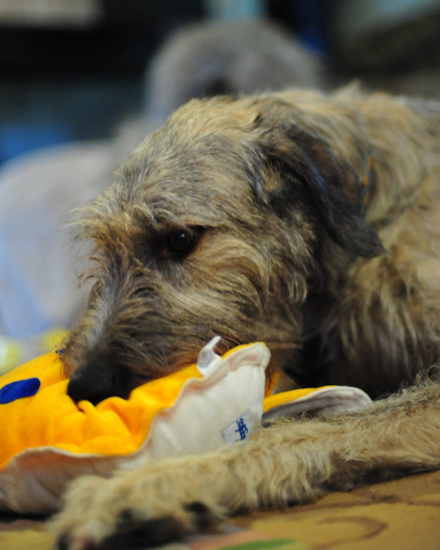
1. Bursa or Hygroma - These are fluid filled sacks which may develop, usually on the elbows, but sometimes on the buttocks or hocks. They usually occur in puppies because they are clumsy and in laying down they often cause repeated trauma to these areas.The body then puts out fluid to act as a cushion. Although unsightly, these are harmless and should go away on their own. It may take a few weeks, or in some cases a few months, but generally they are best left alone. Do not allow anyone to drain them as this just risks the potential for infection and may make the problem much worse. If you cannot encourage the puppy to lay
on padded surfaces, sometimes people have had success by padding the affected part of the anatomy.
You can google "elbow protection for dogs" on the internet to find several options, or you can sew
padding into the sleeve of a sweatshirt and have your dog wear it, if they will.
In any case, this is a condition we usually treat with "watchful neglect".
The picture illustrates a typical elbow bursa on a youngster.
2. Juvenile Vaginitis - It is very common for immature female wolfhounds to develop vaginitis. This is evidenced by a sticky vaginal discharge and often you will notice her doing excess licking at this area. The vaginal opening in a young girl is rather small and as she squats to urinate, she may pick up dirt or debris which then leads to irritation and/or infection. ust keep this area clean. You can clip the hair around the vulva and the rectum to aid in hygiene. If it is bad, you can use a soft rubber pediatric bulb ear syringe and gently
flush out the vagina with a mild douche solution. Doing this every other day for a week or two usually clears up the problem. In any
case, once she matures and goes through her first season, this should no longer be an issue. It is, of course, possible for a vaginal
infection to ascend up the urethra and become a bladder infection or UTI. In my personal experience this has never been an issue, but some people do recommend using cranberry extract as a nutritional supplement to minimize this possibility.
3. Premature Closure of the Distal Ulna or Radius. This can occur in immature dogs with open physes (growth plates). An affected
dog will show signs of intermittent lameness with varying degrees and the lower front limb becomes deformed by bowing. The growth plates are responsible for bone growth in immature dogs, until the growth plates close around one year of age. The growth plates are located at the end of each bone, and immature (open) growth plates are softer than mature bones, so they are more susceptible to injury. The distal (towards the bottom of the bone) growth plate is responsible for 90% of the entire bone’s growth. Causes of
premature closure may include growth plate trauma, abnormal nutrition, hereditary factors, and infection. It is very important to minimize "pounding" type trauma to the front legs. Do not let your wolfhound puppy jump in and out of your vehicle, off your porch or deck, etc. Do not take your puppy jogging or running next to a bike, horse, etc. where he subjects his front legs to continuous pounding. If this problem occurs, it usually starts to be evident around 4-6 months of age. Because of the length of our breed's limbs and the length of growing time, the sooner this conditition is diagnosed and treatment started, the better the outcome. It usually requires surgical correction and sometimes more than one surgery.
4. Fibrocartilaginous Emboli - This is a condition that is very unusual in its presentation in our breed. Most veterinary literature discusses this phenomenon in adult dogs, but in our case it most commonly occurs in 2-5 month old puppies. A normal, happy, healthy puppy is suddenly paralyzed in one or both rear legs with no history of trauma. There is no pain, no fever, no swelling. The puppy may have been trotting across the yard and suddenly goes down and is unable to rise again. I refer you to an excellent article written by Ellen Kroll and published on the Irish Wolfhound Foundation Website.
5. Osteochondritis dissecans, commonly known as OCD, is a disease of the cartilage that can affect various joints in a dog. OCD may affect the shoulder, elbow, knee or hock, although the shoulder is most commonly affected. The symptoms are lameness in the affected limb. Some dogs have a barely noticeable limp and others are unable to bear any weight on the leg. The lameness tends to worsen after periods of exercise and improves after rest. When it affects the shoulder, a shortened forelimb stride may be noted due to reluctance to flex and extend the shoulder joint. Occasionally, the disease will affect both limbs simultaneously and the dog may be reluctant to move. There are currently two ways to treat OCD, conservative medical treatment or surgical removal of the lesion. Conservative treatment may be indicated for dogs that have early mild symptoms of OCD or where a specific lesion cannot be identified on radiographs. Conservative treatment consists of strict rest for 4 to 8 weeks. Leash walking is permitted but no running or playing is allowed. Anti-inflammatories and painkillers such as carprofen (Rimadyl) may be indicated. In addition, the use of glucosamine/chondroitin products has been suggested, yet there are no current studies that confirm their beneficial use in this particular disease. Conservative treatment may be difficult in young, active puppies who may still need to undergo surgery, if the symptoms do not improve. Surgery is indicated in animals that show severe symptoms, in cases where large lesions are identified on radiographs or when conservative treatments fail. The surgery is very straightforward. The affected joint is opened and the offending flap, defect, or joint mouse is removed. The prognosis is generally good when the shoulder joint is affected. With other joints, degenerative joint disease (osteoarthritis) is more common. When the elbow is affected, the OCD may contribute to the development of other abnormalities in that joint.
6. Hip and Elbow Dysplasia - People often assume that all big dogs get hip dysplasia, but may never have heard of elbow dysplasia. Hip dysplasia is a condition that should never be seen in our breed or any of the sighthounds.These dogs were bred for generations based on their ability to gallop. Although the IW is large, he is not heavy compared to some of the other large and giant breed dogs who were bred to do their work at the walk or trot with more concern for chest and shoulder strength and less concern about hindquarter soundness. In more than 50 years in this breed we have never had a case of hip dysplasia. Elbow Dysplasia does occur in IWs and is a general term used to describe one or more of three conditions affecting the bones that form the elbow joint. Breeding stock should always have both hips and elbows x-rayed after they are two years of age and certified free of signs of abnormal joint configuration.
7. Panostetis - Often referred to as Pano. The presenting symptoms include a history of acute sudden lameness not associated with any trauma. It is usually a large breed male dog between the ages of 6 to 18 months. There are periods of lameness lasting from 2 to 3 weeks and it may shift from leg to leg. The most commonly affected bones are the radius, ulna, humerus, femur, and tibia, though the foot and pelvic bones may also be involved. The dog may show a reluctance to walk or exercise. When the affected bones are squeezed, the dog reacts painfully. Occasionally, affected dogs will have a fever, tonsillitis, or an elevated white blood cell count. We have never experienced this problem in any of our young hounds, but if it occurs, be comforted by the fact that it is usually a short lived and self-limiting condition.The treatment involves pain medication and rest.
8. Hypertrophic Osteodystrophy - Another condition we have never seen in any of our IWs, but it certainly could happen and we want owners to be informed. Dogs that are stricken with HOD often show symptoms of mild to moderate painful swelling of the growth plates in the leg bones. It most commonly affects the ends of the radius, ulna, (long bones from the elbow to the wrist) and tibia (long bone from the knee to the hock). The dogs may show lameness and a reluctance to move. They may be lethargic and refuse to eat. A fever may come and go rising as high as 106 degrees. The disease usually affects both legs at the same time. The symptoms may wax and wane and resolve on their own or if the fever is very high for long periods and the bony involvement severe, the dogs may suffer permanent structural damage or even die. The treatment is generally supportive. Since this is a very painful condition
anti-inflammatories and painkillers such as carprofen (Rimadyl) are given. In addition, the animals are usually given a broad-spectrum antibiotic. Strict rest on a comfortable warm bed is recommended. Feeding a nutritious, highly palatable food will help to encourage some dogs to eat. In severe cases steroids may need to be given to control the pain, but because of the possibility of this being a bacterial disease their use may be contraindicated due to their immunosuppressive qualities. Vitamin C is often supplemented though its benefit may be questionable.
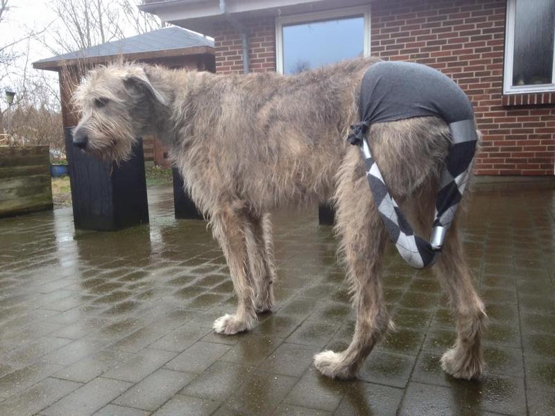
9. Split tail, also called Happy Tail - This is a problem which can occur at any age. Because wolfhounds have long, strong tails and are usually a happy breed, they frequently wag their tails hard and can damage the end by hitting it against a solid surface. When the end of the tail is split it BLEEDS! As they continue to wag it, the blood goes everywhere. In order to control the mess and, more importantly, to prevent repeated trauma and allow healing to occur, many methods have been tried. Most do not work. This is because either the bandage falls off, is put on too tightly and causes further damage which encourages the dog to chew it off and, perhaps, chew at the tail itself, and so forth. One method that seems to work well is to use a pair of old pantyhose. Cut one leg off, put the top of the panty hose on the rear of the dog and slip the tail into the other leg. Then tie the end of the pantyhose leg up onto the waist of the pantyhose. Wrap tape around the tail in two or three places to prevent the tail from slipping out of it's pantyhose sleeve.
Be sure to NOT wrap tape over the injured area. This approach has several advantages;
the fabric allows the wound to breath to encourage healing, it prevents the tail from hitting anything,
it doesn't need to be removed to allow the dog to urinate or defecate and it prevents the dog from
chewing at the end of the tail. The only problem I see would be if the dog chews the whole apparatus
off. Be sure to monitor your dog for a while when you first put this on, so that you can observe
his/her response to it. This picture illustrates the finished product.
10. Liver Shunt - Everyone considering an Irish Wolfhound puppy needs to familiarize\themselves with
the symptoms and testing for Portosystemic Shunts (liver shunt). This is an illness sometimes acquired
in older, or middle aged dogs, but is congenital and genetic in Irish Wolfhound puppies. The two most common types of shunt are intra-hepatic (inside the liver) and extra-hepatic (outside the liver). You can see both types of shunts in Irish Wolfhounds, though by far the most common (and the most difficult to surgically treat) is the intra-hepatic shunt. All wolfhound puppies should be tested by their breeder prior to going to their new homes. If you are thinking of purchasing an IW puppy this should be on your "non-negotiable" list of expectations of your breeder. The earliest puppies can be tested for shunt is 8 weeks of age, but due to how the liver develops I prefer to test later. My personal preference is to test as close to the time the puppies are going home as possible, so I can test at 9 -10
weeks. If puppies are tested prior to 8 weeks there is a greater risk of the test showing the puppy clear of a shunt when in fact he or she isn't, I'll explain this later. If you want to understand why waiting to test is important, you need to understand a little about how the liver works and how shunts develop. The job of the liver is to remove toxins from the body. If it isn't functioning, or functions poorly, toxins build in the body, eventually causing disease processes which will lead to death. If a liver shunt is caught early enough it can be either medically managed or surgically repaired, but even this is still not a guarantee of a positive outcome. The intrahepatic shunt, a blood vessel within the liver itself called the ductus venosus, carries blood from the newborn to the mother bypassing the newborn's liver. This is a natural occurrence in the puppy while in the womb. The mother's liver does the work of removing waste and toxins for her unborn infant; she is functioning for both herself and ALL of her puppies. The puppies' livers are growing and developing during this time and aren't prepared to take on the work load for their little bodies. The need of the mother to process all these toxins is yet another reason why proper feeding and deworming of the bitch is so important. You don't want to make her body work any harder during gestation than it is already. The interior vein (e.g. the ductus venosus) normally closes off at birth, thus allowing the pup's liver to take over the job of removing toxins out of the body. If this fails to happen during development, then a congenital intra-hepatic shunt occurs. If the puppy's body forms an additional and unnecessary blood vessel on the outside of the liver (a congenital vein bypassing the liver on the outside), then this is called an extra-hepatic shunt. If this happens it won't matter if the interior vein closes, you'll still have a shunt moving blood around the liver instead of through it. So, on to the shunts themselves; they are not all created equal. You can have a partial closure of the interior vein where the puppy's liver is doing some of the work of removing toxins, or you can have no closure at all. If you have no closure you will see signs of disease much earlier in the puppy's life, say around 5-7 weeks. If you have a partial closure, or an extra-hepatic shunt that is quite small, you may not see symptoms until a year of age or more. The reason for this is twofold. First, the liver of the puppy is initially quite large in comparison to the size of the puppy's body when it is born. The liver begins growing when the interior vein closes and is often fully formed by 2-4 months of age. This means you would not want to test earlier than 8 weeks when the liver is fully formed. Testing past 8 weeks decreases the risk of false negative results. The reason you sometimes get a negative result with a shunt in place is because the puppy's liver is quite large in relation to its body size and can compensate for a minor shunt for quite some time. This allows a minor shunt to remain "hidden" until the puppy's body has grown to the point where it can no longer compensate. In this scenario the puppy will not test positive until later in life, upwards to a year or even 18 months. A reputable breeder should provide a bile acids panel on all wolfhound puppies prior to sending them to their new homes. The testing may start with a period of fasting, generally overnight, drawing a pre-feeding blood sample on each puppy, feeding a fatty meal, waiting two hours and then drawing a post feeding blood sample on each puppy. The two samples are sent to the lab for testing. The test analyzes the level of bile acids in each sample. If the post feeding sample is overly high a portosystemic shunt is suspected. However, after m,any years of testing, it is now considered normal to screne the litter by just testing a post-feeding sample. Bile acids are removed from the blood in the liver by the liver cells, if these cells aren't functioning as they should the bile acids remain in the blood flow and enter the body blood flow, raising the level of bile acid in the body's blood. Your breeder should be able to provide you with laboratory testing on each puppy showing normal liver values post feeding.
Now, how about some potential adult onset issues:
1. Cardiac issues - Irish Wolfhounds, unlike many smaller breeds, rarely have congenital (present at birth) cardiac issues. The most common cardiac concerns in this breed tend to come on in middle to later life and include Atrial Fibrillation and Dilated Cardiomyopathy. ALL IRISH WOLFHOUNDS, whether they are in show homes, belong to breeders or are someone's beloved companion, should have an annual ECG, beginning at 2-3 years of age, as part of their routine veterinary care. Complete and up to date information on cardiac disease in this breed is available on the Irish Wolfhound Foundation Website at https://www.iwfoundation.org/documents/2016-IWCA_talk_BT_1335379016_8874.pdf
2. Cancer - Cancer can affect dogs of all sizes, genders, purebreds, and mixed breeds. It is estimated that 1 in 3 people and 1 in 3 dogs will be affected by some form of cancer in his/her life. In Irish Wolfhounds, the most commonly seen cancer is osteosarcoma, or bone cancer. It can affect any bone in the body, but most often is seen near the carpus (wrist) in the front legs or the hock (heel) in the hind legs. This form of cancer is usually highly aggressive and tends to metastasize quickly. Although a cure is always the hope, realistically, the goal is to provide palliative care for as long as your companion can enjoy a good quality of life. Amputation of the affected limb does away with the source of pain quickly, but may not be an acceptable option for some cases. Radiation treatment is a new option and is explained well here. http://www.iwfoundation.org/articles_detail.html?item_id=47&year=2014
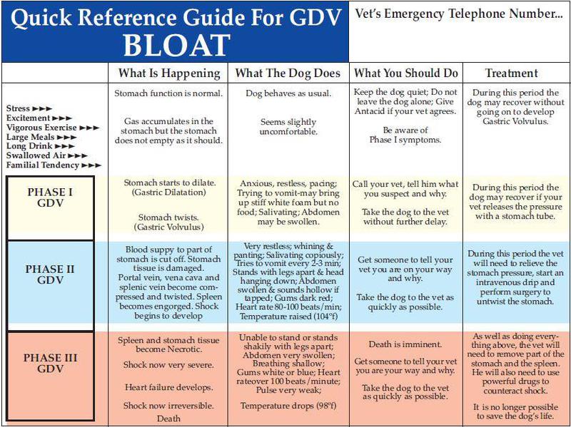
3. Gastric Dilitation/Volvulous (Bloat) - Bloat is a life-threatening emergency that affects dogs in the prime of their life. The mortality rate for gastric dilatation-volvulus (GDV) approaches 50 percent. The keys to survival are early recognition and prompt treatment. Bloat occurs initially when the stomach suddenly distends with gas and fluid.Then the distended stomach rotates on its long axis, a result called volvulus. As the spleen is attached to the wall of the stomach, it rotates as well. Not all cases of gastric dilatation become complicated by volvulus. The stomach can undergo torsion, and twist 180 degrees or less, whereas a full volvulus is a twist of 180-360 degrees or more. The pylorus portion of the stomach is pulled out of position during volvulus, and becomes displaced to the left of the junction of the esophagus and stomach. This squeezes down the duodenum, which prevents fluid and air from escaping the stomach via the pylorus. At the same time, the esophageal-stomach junction is twisted and obstructed, preventing the dog from belching gas and vomiting. Thus, gas and fluid are trapped in the closed-off stomach, which becomes hugely distended as the food ferments. The stomach wall undergoes necrosis once the blood supply is compromised. This sequence of events typically leads rapidly to acute dehydration, bacterial septicemia, circulatory shock, cardiac arrhythmias, gastric perforation, peritonitis and death.
Bloat usually occurs in middle-aged to older dogs, but can occur at any age. There is often a familial or breed association. Large-breed dogs with deep chests are anatomically predisposed, and include: Great Dane, German Shepherd Dog, Saint Bernard, Labrador Retriever, Irish Wolfhound, Great Pyrenees, Boxer, Weimaraner, Old English Sheepdog, Irish Setter, Collie, Bloodhound, Akita, Borzoi, Bullmastiff, Chow Chow, Curly Coated Retriever, Scottish Deerhound, German Longhaired Pointer, Grand Bleu de Gastogne, Mastiff, Neopolitan Mastiff, Newfoundland, Bernese Mountain Dog, Doberman Pinscher, Gordon Setter, Greater Swiss Mountain Dog, Greyhound, Rhodesian Ridgeback, and Standard Poodle. Chinese Shar-Pei and the Basset Hound have the highest incidence among mid-size dogs. Small dogs are rarely affected, with the exception of the Dachshund varieties, who are also deep-chested.
The clinical history of bloat cases often includes a sudden onset in a healthy, active dog. He may have just eaten a large meal, exercised vigorously before or after eating, or had a large amount of liquid immediately after eating.
The classic signs of bloat are restlessness and pacing, salivation, retching, frequent attempts to vomit, and an enlarging abdomen. The dog may whine or groan when you press on his belly. Thumping on the abdomen produces a hollow sound, like a kettle drum.
Unfortunately, not all cases of bloat present with classical signs. In the early stage, the dog may not appear distended, although the abdomen usually feels slightly tight. The dog appears lethargic, is obviously uncomfortable, walks stiff-legged, and may hang his head. At this stage, one cannot distinguish dilatation from volvulus.
Late signs of impending shock are obvious, including pale gums and tongue, delayed capillary refill time, rapid heart rate, weak pulse, rapid and labored breathing, weakness, and collapse.
Whenever there is even a slight suspicion of bloat, take the dog immediately to a veterinary hospital. If is too late for that, be brave and take a clean, sharp knife and insert it quickly into the bloated side of the dog to let the trapped gas escaped from his distended stomach. This emergency procedure has saved lives – then immediately go to the closest veterinary hospital.
At the veterinary clinic, gastric dilatation without torsion or volvulus is relieved by passing a long rubber or plastic tube through the dog’s mouth into the stomach. As the tube enters the dog’s stomach, a rush of air and fluid from the tube will bring relief. The stomach is then washed out. The dog should not eat or drink for the next 36 hours, and will need to be supported with intravenous fluids. If symptoms do not return, the regular diet can be gradually restored.
In contrast to simple dilatation, abdominal x-rays reveal a normal gas pattern but an excess amount of it in the stomach. With volvulus, however, x-rays have a “double bubble” gas pattern, with gas appearing in two sections separated by the twisted tissue.
In volvulus cases, emergency surgery is required as soon as the dog is stable enough for anesthesia. At surgery, the stomach and spleen are re-positioned, unless there is necrosis which requires removing the spleen and even a portion of the stomach.
The risk of recurrence for dogs responding to nonsurgical treatment is 70%. For that reason, dog caregivers and breeders may elect to have the dog’s stomach tacked down to the rib cage to prevent volvulus (gastropexy). Other preventives measures are critical, and include:
• Divide the day’s ration into 2-3 equal meals, spaced well apart.
• Do not use a raised food bowl.
• Avoid dry dog food high in fat (listed in the first 4 ingredients on the label).
• Avoid foods that contain citric acid.
• Restrict access to water for 1 hour before and after meals.
• Never let the dog drink a large amount of water all at once.
• Avoid strenuous exercise on a full stomach.
In addition to this overview, it is my personal opinion that stress plays a major role in the
development of bloat. For this reason, I strongly recommend do as much socialization as
possible with your dogs, starting as soon as you get your puppy. The goal is to help your dog
develop a strong self confidence to minimize stress in unusual situations. I normally do not
recommend gastropexy (tacking the stomach to prevent torsion) as an elective surgery.
My feeling is why subject your pet to an elective procedure that most likely will never be needed.
The vast majority of Irish Wolfhounds never have a bloat episode in their lives. However, if your dog undergoes any abdominal surgery; ie. spay, C-section, laparotomy or most definitely in the case of gastric torsion, then do have the stomach tacked down to minimize future problems.
4. Pneumonia - Pneumonia in the Irish Wolfhound can become life-threatening so quickly that it is imperative for owners and their veterinarians to recognize this disease and its serious nature. A positive outcome depends upon prompt treatment. Diagnosing pneumonia is not always easy, because wolfhounds are notoriously ‘stoic’ and may not act particularly ill early in the disease, and because the first symptoms can be somewhat vague and may vary from case to case. Because of these factors, a dog can be very ill indeed before it becomes obvious that something is wrong.
Most cases of pneumonia in dogs are related to some ‘predisposing factor. Typical predisposing factors include other illnesses (kennel cough complex or distemper virus, for example), aspiration into the lungs (most commonly in anesthetized dogs or dogs who may have swallowing or esophagus problems, or laryngeal paralysis), or systemic infection (sepsis). Nonetheless, many wolfhound owners have had the experience of a hound becoming desperately ill with pneumonia without any obvious predisposing factor. A hound can seem perfectly fine and then, within a matter of hours, be seriously ill. Survival can depend upon rapid treatment with antibiotics, so owners and vets must be sensitized to the possibility that a sick hound has pneumonia even if none of the ordinary predisposing factors are present. IWs may very well have an underlying susceptibility which is currently under investigation by researchers at the University of Pennsylvania, so tell your veterinarian to keep that fact in mind if he seems unwilling to initiate treatment.
A new strain of the canine influenza virus has spread from greyhound racing kennels in Florida to dog shows in the southeast. Since this is a new strain, no dogs have immunity. Most dogs experience only mild upper respiratory symptoms, but some do get pneumonia, with a death rate between 1% and 5%, so beware.
Symptoms - If your dog develops pneumonia, the first two things you may notice are lethargy and loss of appetite. Some hounds run fevers, often very high, while others do not. Probably the most universal symptom in the IW is the typical "pneumonia posture" of extended neck and head, which the wolfhound assumes to make breathing easier and take in more air. The neck and head are lowered, level with the back, and the head is held straight out. Usually a wolfhound with pneumonia will not be comfortable lying on its side. Most wolfhounds don’t have nasal secretions with pneumonia, although some may. Some may also display rapid or difficult breathing early on. In some cases a hound will appear to have abdominal distress. A dog displaying signs like these needs to see a vet as quickly as possible. Suspected pneumonia in an Irish Wolfhound can never be taken lightly.
Diagnostic Tests - X-rays may or may not help your vet diagnose pneumonia. Clinical evidence shows that a wolfhound can be gravely ill and in danger even before changes in the lung tissue can be visualized on an X-ray, so a clear chest X-ray does not rule out pneumonia. Auscultation (listening to the lungs) is frequently, but not always, helpful. The lungs can be cultured using a tracheal wash if the hound is stable enough to undergo light sedation. This procedure is valuable because results can help guide treatment choice, especially if the IW does not respond to initial antibiotic therapy. However, your vet must initiate antibiotic therapy before results are obtained. Fungal infections display a typical pattern on chest x-rays, but secondary bacterial infection is usually present as well. If an IW does not improve, or relapses when antibiotics are discontinued, it is prudent to rule out fungal infection. Depending on your area, blastomycosis and valley fever are two fungal diseases which can be devastating if left untreated.
Dr. Phil Padrid, a specialist in respiratory disease and a regional medical director of VCA Animal Hospitals, also recommends monitoring blood gases (or pulse oximetry, if blood gas monitoring is not available) as a more reliable tool than x-ray films for assessing a dog’s condition.
Infectious Agents and Antibiotics - Wolfhounds with pneumonia may have a number of different types of bacteria in their lungs: streptococci, staphylococci, e. coli, etc, may be seen alone or in combination. Mycoplasma and fungal infections are also possibilities. Dr. Padrid suggests it is not a good idea to discount anaerobic bacteria such as e. coli and klebsiella when choosing antibiotic therapy. He recommends 3 weeks of Baytril and Antirobe be given together to cover a broad range of both aerobic and anaerobic, gram-positive and -negative, pathogens. Dr. Margret Casal, University of Pennsylvania Veterinary School, suggests Zithromax, an antibiotic belonging to the class called macrolides, may be a good first choice for pneumonia, because it is effective against a variety of organisms and can be given in one single dose per day. In addition, Zithromax remains at therapeutic levels in the bloodstream for up to ten days after treatment is discontinued.
There are a number of antibiotics in several different classes (including macrolides, fluoroquinolones, cephalosporins) that are available for use in dogs with pneumonia. Among the drugs which are frequently prescribed, Baytril and Zeniquin ( fluoroquinolones), Keflex and Naxcel (first- and second-generation cephalosporins) are a few of the other drugs which have proven successful in some individual cases of IW pneumonia. Whatever your veterinarian selects, if your IW does not begin to improve rapidly, your veterinarian must consider either adding a second drug or switching drugs.
Additional Therapy - In addition to antibiotic treatment, dogs with pneumonia need to be kept well-hydrated. This may require IV or subcutaneous fluids. Sometimes expectorant medicines (like Mucinex, available over the counter in your drug store) help clear the lungs, but cough suppressants should be avoided. Coupage, or rapidly tapping the chest wall, is excellent for helping your IW clear his lungs. Coupage should be performed three or four times each day.
It is also important for your IW to have light exercise; (especially important if your dog requires hospitalization, so make sure the caregiver knows this!) but remember that lung capacity is diminished. Just make sure your IW is not outside any longer than necessary in rainy or cold weather. You and your vet should carefully monitor your hound’s progress. Again, pneumonia in Irish Wolfhounds is a very serious illness. Failing to treat promptly and aggressively can be fatal!
For additional information regarding pneumonia in Irish Wolfhounds visit the UK Irish Wolfhound Health Group at